What Does A Cavity Look Like?
What does a cavity look like is the first question people google when they feel the twinge of tooth pain. The answer isn’t pretty but it’s important that we take a look. Cavities, also known as dental caries or tooth decay, are damaged areas in the hard surface of your teeth that develop into tiny openings or holes. They are among the world’s most common health problems, affecting people of all ages. Cavities are caused by a combination of factors, including bacteria in your mouth, frequent snacking, sipping sugary drinks, and not cleaning your teeth well. Recognizing a cavity early can save not only the tooth but also save one from undergoing extensive dental procedures.

Appearances Matter: What Exactly Does A Cavity Look Like?
What does a cavity really look like? Cavities can start as tiny spots that slowly turn into large holes if left untreated. By understanding the stages of tooth decay, you can catch cavities early before they cause pain or lasting damage.
In the beginning, you may notice small white, tan or brown spots appear on your teeth near the chewing surfaces or between teeth. These early signs of decay are often painless. The spots form when acids from foods or bacteria dissolve minerals from your tooth enamel. If the demineralization continues, the spots turn darker as the enamel decays further. Eventually, this creates a small hole or weakened area in the hard, outer enamel layer.
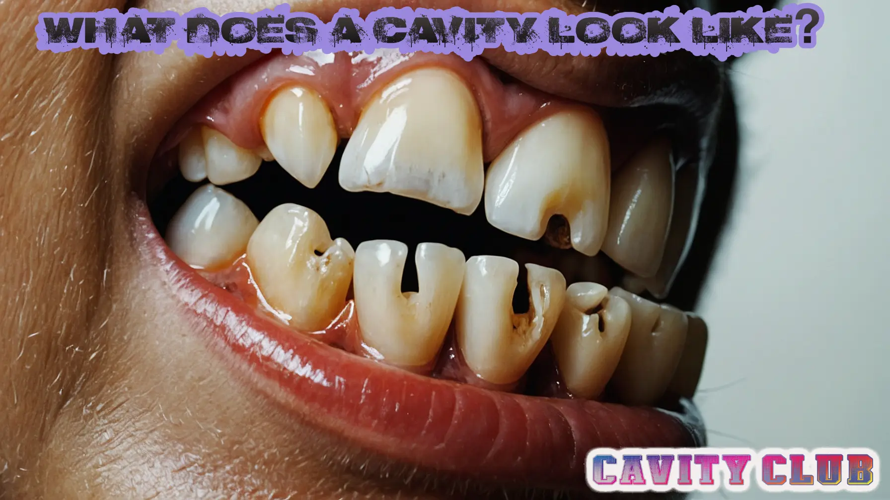
At this point, a small cavity has formed. Tiny cavities may look like pinprick dents or holes surrounded by discolored enamel. You may be able to see a dark spot indicating the hole. Small cavities can be easier to treat by brushing with fluoride toothpaste, using dental sealants or applying topical fluoride at the dentist.
As the decay spreads through enamel and reaches the softer dentin layer under the enamel, the cavity becomes larger and deeper. The hole appears more pronounced, with visibly missing enamel. You may notice tooth discoloration around the hole as the enamel weakens. The cavity edges appear tan, dark brown or black. Looking inside the hole, the dentin has a yellowish hue.
Larger cavities can be round, oval or angular shaped with depths ranging from 1 to 6 millimeters. The holes become big enough to trap food easily. You may experience some pain from hot, cold or sweet foods as the inner tooth layers are exposed. But it still may not cause severe constant pain.
At this moderate decay stage, fillings become necessary to restore the tooth structure. If cavities grow untreated, they can reach the inner soft pulp tissue, become severely infected, and cause abscesses or tooth loss. You would experience throbbing pain at this point.
The key is to not ignore small spots and holes in your teeth, hoping they will go away. Have your dentist examine any discolored areas for early signs of tooth decay. Catching cavities when they are small prevents worsening decay, so you can maintain strong, healthy teeth for life.
What Does A Cavity Look Like At Each Stage Of Formation?
Cavities start with demineralization as acids weaken the enamel. This leads to enamel decay, where small holes form in the enamel surface. The cavity then spreads to dentin decay, with larger holes forming in the softer dentin underneath. Finally, in pulp involvement, the decay reaches the pulp, causing severe pain and requiring major treatment. Catching cavities early through dental visits allows treatment before extensive enamel and dentin decay, preventing pulp damage.

Demineralization
In the beginning stage of cavity formation, you may notice small white, tan or brown spots appearing on your teeth. This is known as tooth demineralization, where acids from food and bacteria dissolve minerals like calcium from the enamel. The spots indicate areas where the enamel has started to weaken. At this early stage, the spots are often painless and may come and go. But they show the first signs of tooth decay, so see your dentist to treat demineralization before it can worsen into a cavity.
Enamel Decay
As tooth decay worsens, tiny holes or pits start to form in the enamel surface – this is called enamel decay. The small cavities may appear as pinprick dents or holes surrounded by darkened, discolored enamel. You can often see a brown or black dot indicating the location of the hole. At this point, the cavity has eaten through the hard outer enamel layer but is still small. Getting dental sealants or fluoride therapy now can remineralize these budding cavities before they grow larger and impact inner tooth layers.
Dentin Decay
As a cavity burrows past the enamel and reaches the softer dentin layer, you’ll notice a larger, more pronounced hole in your tooth. The cavity edges appear dark brown or black with visibly missing enamel around it. Inside, the exposed dentin has a yellowish color compared to the white enamel. The hole may be round, oval or angular in shape. As more dentin is impacted, you may feel some temperature sensitivity or pain from sweets as deeper tooth nerves become irritated. At this point, you’ll likely need a filling to treat the worsening decay.
Pulp Involvement
In the most advanced stage of tooth decay, known as pulp involvement, the cavity has burrowed through dentin and reached the pulp tissue inside the tooth. The soft, reddish-pink pulp contains nerves and blood vessels essential for the tooth’s health. Exposure leads to inflammation or infection, causing throbbing, severe tooth pain. Pus may drain from the gum near the infected tooth. At this critical point, you need a root canal or the tooth will likely be lost. Don’t delay treatment if you have extreme, constant pain indicating pulp damage.

What Does A Cavity Look Like In An X Ray?
When you get dental x-rays taken, they provide a unique glimpse into what’s happening inside the hard tooth structure. X-rays pass through the enamel and dentin layers to produce an image detailing both the surface and inner areas of each tooth. One key thing x-rays can reveal is the development of cavities. Before a cavity forms an externally visible hole, it often starts as a small zone of demineralization deep in the layers of enamel or dentin. An x-ray is able to detect these budding areas of decay.
On an x-ray image, healthy tooth structure appears radiopaque, meaning solid white, due to the dense mineral content blocking most x-rays. In contrast, decayed regions allow more x-ray penetration, causing them to look dark or transparent. The level of darkness corresponds to the extent of mineral loss. Incipient cavities confined to enamel may look faint, with light gray shading demonstrating surface lesions. However, more advanced cavities burrowing deep into dentin appear as markedly darker spots with crisp borders. The varying shapes – round, oval, or angular – depict the shape of the hollowed region eating away healthy tooth layers.
In the worst cases, cavities can reach the inner pulp tissue. This shows on x-rays as darkened areas spreading across the entire inner tooth width, signifying dying or infected pulp. However, most cavities are caught sooner in routine checkup x-rays. Being able to visualize small areas of tooth decay when they are still reversible is the best early diagnostic tool. This allows preventative treatment with fluoride or sealants before cavities require fillings or worsen to cause damage beyond the power of x-rays to describe in shaded pictures alone.
Make Sure You Never Have To Look At Cavities
The National Institute of Dental & Craniofacial Health recommends thrice-daily brushing, with flouride if it’s not in your city water supply, daily flossing, limiting sweets and eating well, and regular trips to the dentist for check-ups.
But If You Do, Make A Cavity Disappear Like:
Treating cavities ranges from preventative care for initial decay to more invasive methods for advanced decay. The goal is to save the tooth structure and prevent further damage. The type and how much a cavity filling costs will depend on several factors such as the size, depth and location of your cavity, your dentist will recommend the most appropriate treatment method.
If caught early when white, brown or black spots signal demineralization below the enamel surface, topical fluoride or dental sealants can help remineralize these areas. Fluoride therapy replaces missing minerals, especially calcium and phosphate, needed to rebuild and restore enamel. Synthetic resins used as dental sealants also halt the decay process by adhering to pitted areas vulnerable to food and bacteria. These treatments are painless and aim to prevent full cavity formation.
For small to mid-sized cavities confined to a single tooth surface with minimal discomfort, traditional amalgam or composite resin fillings allow repair of holes or cracks from decay. The dentist first numbs the area then uses a drill to remove bacteria and decayed material. This creates a clean inner cavity space to fill and rebuild with amalgam or a tooth-colored resin bonded to the surface to completely seal out food, drink and bacteria. Though impermanent with typical lifetimes under 10 years, fillings adequately restore teeth at moderate cost.
Larger cavities, especially a cavity between teeth or approaching the pulp space inside can be more difficult to treat. Onlays or inlays serve as customized indirect fillings, shaped and colored in a dental lab before adhering over deep decay. Crowns fully encase damaged teeth to prevent fracturing and provide support. Root canals remove infected pulp then tightly seal and restore the deadened tooth. Lastly, extraction safely removes non-restorable severely decayed teeth. Implants may offer replacements. Listen to your dentist’s advice on which solution best resolves your specific dental cavities. Catching decay early allows simpler treatments for long-term tooth preservation.
Frequently Asked Questions
A small cavity may appear as a tiny hole or pit in the enamel of a tooth, often with brownish discoloration. The cavity is usually painless in early stages.
A deep cavity may look like a large hole in the tooth, sometimes with visible dentin underneath. It may cause tooth sensitivity or pain when eating sweets or cold/hot foods.
On an X-ray, a cavity appears as a dark spot or hole in the white enamel of the tooth. The cavity may appear minor or more advanced depending on the extent of decay.
No, cavities can vary in shape, size and location depending on factors like where they form and how advanced the decay is. Shallow cavities may look like small discolored spots while deeper cavities look like holes.
Early childhood cavities often start as pale white or yellow spots on the front and back teeth. As decay worsens, they turn brown and form holes in the teeth.